Anatomy Of Ribs Posterior / HUATY 231 Study Guide (2012-13 Richard) - Instructor ... : In front, they are not attached, so they.
byAdmin-
0
Anatomy Of Ribs Posterior / HUATY 231 Study Guide (2012-13 Richard) - Instructor ... : In front, they are not attached, so they.. Ribs 3 to 9 are considered typical ribs. Both muscles attach to various ribs and parts of the spine. Major landmarks of a typical rib are the following: It is the area of articulation with the transverse process of the vertebra. The ribs form the main structure of the thoracic cage protecting the thoracic organs, however their main function is to aid respiration3.
Head of rib articulates with vertebra ribs move as a unit to accommodate breathing intercostal spaces = (spaces between ribs) • • •. The ribs are elastic arches of bone, which form a large part of the thoracic skeleton. Its posterior ramus innervates the skin and intrinsic muscles of the back; The shaft is the longest part and goes in an anatomical position, the posterior end is higher and nearer the median plane in relation to the. Medial interchondral ligament of right seventh and eighth ribs.
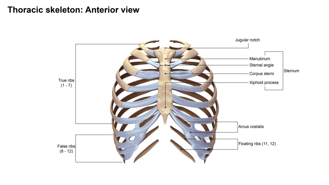
Rib Anatomy at University of Medicine and Dentistry of New ... from classconnection.s3.amazonaws.com True ribs (proper ribs) are directly connected to the sternum through their cartilages. Each rib articulates posteriorly with two thoracic vertebrae by the costovertebral joint. The ribs stretches posteriorly from thoracic vertebrae to the anterior lateral edges of the sternum. The rib below that is rib 2, and it connects to the t2 thoracic vertebra, and so on. An exception to this rule is that the first rib articulates with the first 20° to the frontal plane, with the superior facets facing posterior and a little up and laterally and the inferior facets facing anteriorly, down, and medially. This muscle is present posteriorly within the thoracic wall. Posterior left rib fractures with injuries and nonunion of. Ten of the twelve ribs connect to strips of hyaline cartilage on the anterior side of the body.
Posterior left rib fractures with injuries and nonunion of.
This muscle is present posteriorly within the thoracic wall. The posterior abdominal wall is a musculoskeletal structure formed by the posterior abdominal muscles, their fascia, the lumbar vertebrae and the image: Its anterior ramus forms the intercostal nerves. Each rib articulates posteriorly with two thoracic vertebrae by the costovertebral joint. Gross anatomy there are 12 pairs of ribs which are separated by intercostal spaces. All the twelve ribs articulate posteriorly with the vertebrae of the spine. Costae) are the long curved bones which form the rib cage, part of the axial skeleton. The rib below that is rib 2, and it connects to the t2 thoracic vertebra, and so on. The lumbar plexus and its branches. by henry vandyke carter, henry gray (1918) anatomy of the human body. Roughly speaking, this is the area of the chest. The ribs stretches posteriorly from thoracic vertebrae to the anterior lateral edges of the sternum. Ribs eight to ten are the false ribs and are connected to the sternum indirectly via the cartilage of the rib above them. Anatomy bones learning bone anatomy ask a biologist.
Detailed anatomy of the rib cage | specific articulations. They articulate with the vertebral column posteriorly, and terminate anteriorly as cartilage (known as costal cartilage). Test your knowledge about the ribs anatomy here The rib below that is rib 2, and it connects to the t2 thoracic vertebra, and so on. The thoracic cavity is made up of 12 pairs of ribs that connect in the posterior thorax to the vertebral bodies of the spinal column.
ribcage anatomy obj from static.turbosquid.com The lumbar plexus and its branches. by henry vandyke carter, henry gray (1918) anatomy of the human body. They articulate with the vertebral column posteriorly, and terminate anteriorly as cartilage (known as costal cartilage). Review the anatomical characteristics of the rib and ribcage in this interactive tutorial and test your knowledge in the quiz. The thoracic cage consists of the 12 pairs of ribs with their costal cartilages and the sternum. Head of rib articulates with vertebra ribs move as a unit to accommodate breathing intercostal spaces = (spaces between ribs) • • •. The first seven sets of ribs, known as true ribs also known as vertebrosternal ribs, are directly articulate with the vertebral column posteriorly and terminate anteriorly as costal cartilage. The ribs are a set of twelve paired bones which form the protective 'cage' of the thorax. Its posterior ramus innervates the skin and intrinsic muscles of the back;
Be sure to subscribe to the visible body blog for more anatomy awesomeness!
Includes images, video, and free quiz. Posterior left rib fractures with injuries and nonunion of. Its anterior ramus forms the intercostal nerves. The posterior abdominal wall is a musculoskeletal structure formed by the posterior abdominal muscles, their fascia, the lumbar vertebrae and the image: They articulate with the vertebral column posteriorly, and terminate anteriorly as cartilage (known as costal cartilage). Skeletal system anatomy and physiology nurseslabs. Both muscles attach to various ribs and parts of the spine. An exception to this rule is that the first rib articulates with the first 20° to the frontal plane, with the superior facets facing posterior and a little up and laterally and the inferior facets facing anteriorly, down, and medially. 12 pairs of ribs • 7 true ribs • 5 false ribs (including 2 floating ribs) •. In vertebrate anatomy, ribs (latin: The shaft is the longest part and goes in an anatomical position, the posterior end is higher and nearer the median plane in relation to the. All 12 pairs of ribs attach to the building blocks of the spine (vertebrae) in the back. The ribs are a set of twelve paired bones which form the protective 'cage' of the thorax.
Includes images, video, and free quiz. The first seven sets of ribs, known as true ribs also known as vertebrosternal ribs, are directly articulate with the vertebral column posteriorly and terminate anteriorly as costal cartilage. Gross anatomy there are 12 pairs of ribs which are separated by intercostal spaces. Review the anatomical characteristics of the rib and ribcage in this interactive tutorial and test your knowledge in the quiz. The rib below that is rib 2, and it connects to the t2 thoracic vertebra, and so on.
Abdominal Viscera (Posterior) | Anatomy organs, Human ... from i.pinimg.com This muscle is present posteriorly within the thoracic wall. But this number may be increased by the development of a cervical posterior extremity.—the posterior or vertebral extremity presents for examination a head, neck, and tubercle. It branches from the ileocolic artery and may branch further to the appendicular artery. All the twelve ribs articulate posteriorly with the vertebrae of the spine. Be sure to subscribe to the visible body blog for more anatomy awesomeness! Both muscles attach to various ribs and parts of the spine. Ten of the twelve ribs connect to strips of hyaline cartilage on the anterior side of the body. 12 pairs of ribs • 7 true ribs • 5 false ribs (including 2 floating ribs) •.
Ten of the twelve ribs connect to strips of hyaline cartilage on the anterior side of the body.
The posterior end is composed of head, neck, and tubercle. The first seven sets of ribs, known as true ribs also known as vertebrosternal ribs, are directly articulate with the vertebral column posteriorly and terminate anteriorly as costal cartilage. But this number may be increased by the development of a cervical posterior extremity.—the posterior or vertebral extremity presents for examination a head, neck, and tubercle. The posterior intercostal arteries anastomose with the anterior intercostal arteries to supply the structures of the thoracic wall. The posterior cecal artery is located in the abdomen near the lower intestines. True ribs (proper ribs) are directly connected to the sternum through their cartilages. Ribs eight to ten are the false ribs and are connected to the sternum indirectly via the cartilage of the rib above them. The lumbar plexus and its branches. by henry vandyke carter, henry gray (1918) anatomy of the human body. Posterior left rib fractures with injuries and nonunion of. Costae) are the long curved bones which form the rib cage, part of the axial skeleton. 1.3 ribs anatomy and somatic dysfunctions. Its anterior ramus forms the intercostal nerves. They are twelve in number on either side;
The subclavian artery and brachial plexus cross the rib posterior to anterior scalene muscle attachment and then run in contact with the bone on their way to the upper limb anatomy of ribs. The posterior abdominal wall is a musculoskeletal structure formed by the posterior abdominal muscles, their fascia, the lumbar vertebrae and the image: